
Panel and add-ons at a glance
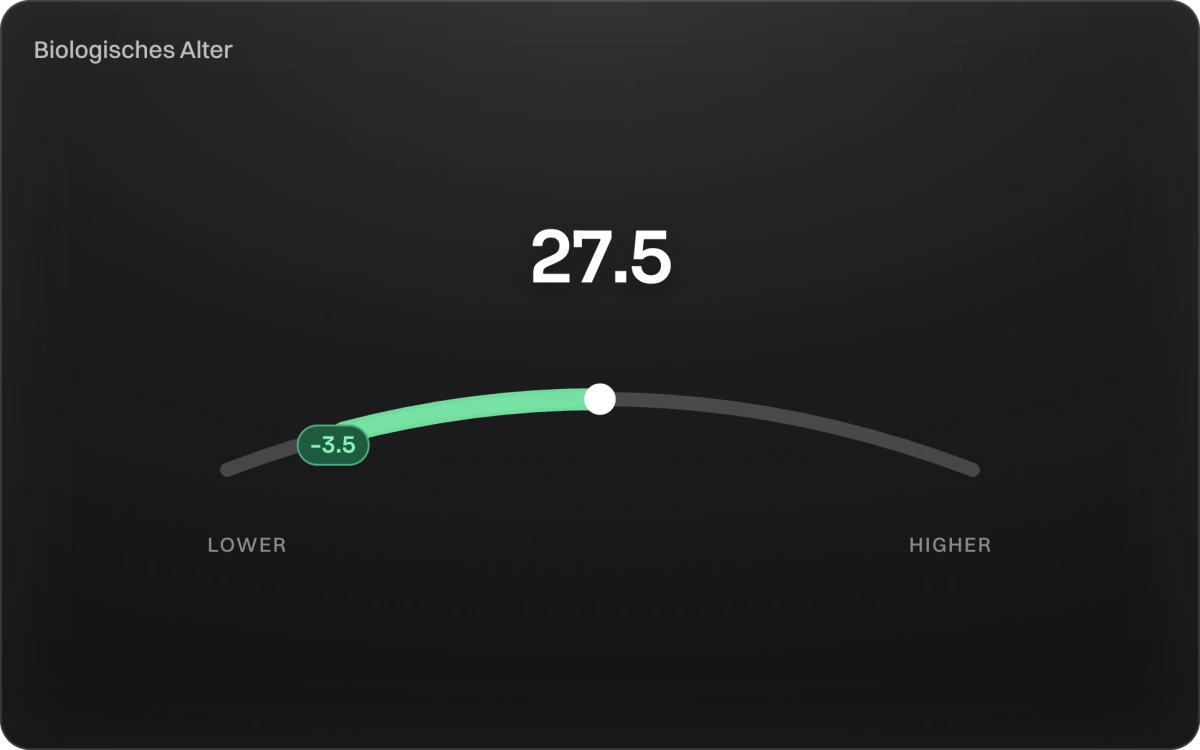
Your biological age
from 9 routine valuesYour biological age shows how fast your body is ageing, regardless of the calendar. NOA calculates it as PhenoAge after Levine, from nine routine values in your analysis.
The biological age is for information only. It is an approximation of your current ageing status, not a diagnostic instrument.
What are biomarkers?
Biomarkers are measurable values in your blood that show how well your body is working. Every result is a glimpse into your health, from heart and metabolism through hormones and nutrients to inflammation and stress.
Measure these values over time to spot change early, follow your progress and steer where it matters.
We group the NOA Core values into 5 areas — they are part of every NOA analysis. Below them you will find 4 add-on panels you can book individually: Performance, Hormone balance, Lp(a) and Omegabalance.
46 values additionally carry a Retest. You can add the retest upgrade; it repeats the check after about six months, so you see the direction.

Heart & metabolism
Blood lipids, blood sugar and fatty acids: the values that set your cardiovascular risk and your energy metabolism.
Total Cholesterol Core measured Retest
Total cholesterol measures the overall amount of cholesterol in the blood.
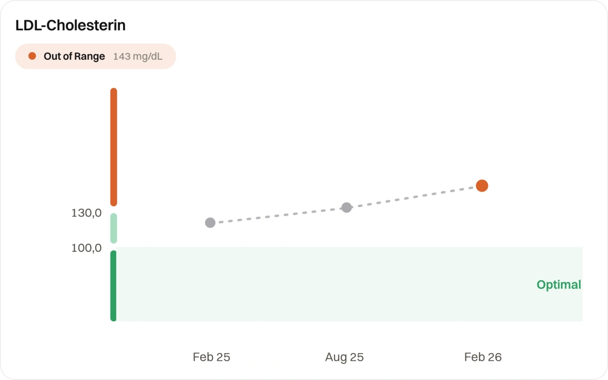
LDL Cholesterol Core measured Retest
LDL cholesterol measures the amount of cholesterol carried by low-density lipoprotein (LDL) particles in the blood.
HDL Cholesterol Core measured Retest
HDL cholesterol measures the amount of cholesterol carried by high-density lipoprotein (HDL) particles in the blood.
Triglycerides Core measured Retest
Triglycerides measure the amount of fat (lipids) circulating in the blood.
Apolipoprotein B Core measured Retest
Apolipoprotein B (ApoB) measures the number of atherogenic cholesterol-carrying particles in the blood.
Non-HDL Cholesterol Core calculated Retest
Non-HDL cholesterol is calculated as total cholesterol minus HDL cholesterol.
Remnant Cholesterol Core calculated
Remnant cholesterol is the cholesterol carried in triglyceride-rich lipoproteins and their breakdown products, that is VLDL, IDL and…
VLDL cholesterol Core calculated
VLDL stands for very low density lipoprotein. These particles carry mainly triglycerides from the liver into tissue. The value on a report…
Triglyceride/HDL Ratio Core calculated Retest
The TG:HDL ratio divides fasting triglycerides by HDL cholesterol.
Atherogenic Index Core calculated
AIP is a calculated index expressed as the logarithm (log) of the ratio of fasting triglycerides to HDL cholesterol.
Atherogenic Coefficient Core calculated
The atherogenic coefficient is calculated as (total cholesterol − HDL) / HDL and equals the ratio of all “non-protective” cholesterol (all…
Total Cholesterol / HDL Ratio Core calculated Retest
The total cholesterol to HDL ratio divides total cholesterol by HDL cholesterol, setting all the cholesterol circulating in the blood…
LDL Cholesterol / Apolipoprotein B Ratio Core calculated
Because each LDL particle carries exactly one molecule of apolipoprotein B, the ratio of LDL cholesterol to ApoB estimates how much…
Triglyceride / Apolipoprotein B Ratio Core calculated
This ratio describes the average triglyceride content per ApoB particle and therefore sheds light on lipoprotein composition , how…
Triglyceride/Cholesterol Ratio Core calculated
The triglyceride/cholesterol ratio is calculated by dividing the triglyceride concentration by total cholesterol.
Neutrophils / HDL Ratio (NHR) Core calculated
The NHR relates neutrophil granulocytes , key cells of the acute, non-specific immune response , to HDL cholesterol with its…
TyG Core calculated Retest
The triglyceride-glucose index (TyG) combines fasting triglycerides and glucose in a logarithmic formula to estimate insulin resistance…
GGT / HDL Cholesterol Ratio Core calculated
This ratio relates gamma-glutamyltransferase (GGT) , a liver enzyme that also reflects oxidative stress , to HDL cholesterol, which has…
Uric Acid / HDL Cholesterol Ratio Core calculated
The uric acid/HDL ratio (UHR) connects uric acid , an end product of purine metabolism associated with oxidative stress and inflammation , …
hs-CRP / HDL Ratio Core calculated
The ratio relates high-sensitivity C-reactive protein (hs-CRP) , a sensitive marker of low-grade inflammation , to anti-inflammatory HDL…
Fasting insulin Core measured
Insulin is a hormone produced by the pancreas that helps regulate blood glucose by enabling glucose to enter cells for energy use or…
HOMA-IR (insulin resistance) Core calculated
HOMA-IR (Homeostatic Model Assessment of Insulin Resistance) is a mathematical index calculated from a single set of fasting glucose and…
Glucose Core measured Retest
Fasting glucose measures the level of glucose (sugar) in the blood after an overnight fast, typically of at least 8 hours.
Hemoglobin A1c (HbA1c) Core measured Retest
Your average blood sugar over the last two to three months, a longer-term view than a single reading. It's the standard way to catch blood sugar drifting up early.
Glucose, estimated average (eAG) Core calculated
Estimated average glucose (eAG) is a mathematical conversion of the HbA1c percentage into average blood glucose units (mg/dL or mmol/L), …
METS-IR Core calculated
METS-IR stands for Metabolic Score for Insulin Resistance. It estimates insulin sensitivity from fasting glucose, triglycerides, HDL…
SPISE Core calculated
SPISE stands for Single Point Insulin Sensitivity Estimator. It estimates insulin sensitivity from three values out of a single blood…
HOMA-Beta (beta-cell function) Core calculated
HOMA-Beta estimates from fasting insulin and fasting glucose how much insulin the pancreatic beta cells still supply. It is the counterpart…
QUICKI (insulin sensitivity index) Core calculated
QUICKI (Quantitative Insulin Sensitivity Check Index) is a mathematical index calculated from fasting insulin and fasting glucose: QUICKI =…
High-Sensitivity C-Reactive Protein (hs-CRP) Core measured Retest
High-sensitivity C-reactive protein (hs-CRP) is a laboratory measurement of C-reactive protein (CRP), an acute-phase protein made mainly by…
hs-CRP / Albumin Ratio (CAR) Core calculated
The C-reactive protein/albumin ratio (CAR) sets a positive acute-phase protein (hs-CRP, rises with inflammation) against a negative one…
hs-CRP / Ferritin Ratio Core calculated
This ratio relates two markers that both rise with inflammation: rapidly responding hs-CRP and more slowly responding ferritin.
Ferritin / Albumin Ratio Core calculated
The ratio connects ferritin , a storage marker that is also an inflammation marker (positive acute-phase protein) , with albumin, which…
PhenoAge (biological age, Levine) Core calculated Retest
PhenoAge is a score published in 2018 by Morgan Levine and colleagues that derives a calculated biological age from nine routine values:…

Hormones
Thyroid, sex hormones and the stress axis: the control system behind energy, mood and recovery.
Thyroid-Stimulating Hormone (TSH) Core measured Retest
Thyroid-stimulating hormone (TSH) is produced by the pituitary gland in the brain and functions as the master regulator of the thyroid , it…
Cortisol (morning value) Core measured
Cortisol is the adrenal gland's main stress hormone. It follows a daily rhythm: highest shortly after waking, falling through the day,…
Sex Hormone-Binding Globulin (SHBG) Core measured
Sex hormone-binding globulin (SHBG) is a glycoprotein produced by the liver that binds tightly to testosterone and estradiol, keeping them…
Total Testosterone Core measured
Total testosterone measures all testosterone circulating in the blood, both the small free (unbound) fraction and the larger fractions bound…
Free Testosterone (Vermeulen) Core calculated
Free testosterone by Vermeulen is calculated from total testosterone, SHBG and albumin. In blood, most testosterone is tightly bound to…
Free Testosterone Index (FTI) Core calculated
The Free Androgen Index (FAI) is a calculated ratio of total testosterone to sex hormone-binding globulin (SHBG), multiplied by 100.

Vitamins, minerals & iron
The building blocks of energy, immune system and muscle function, measured in serum and inside the cell.
Sodium Core measured Retest
Sodium is the dominant positively charged ion in the extracellular fluid , the blood and the fluid bathing cells and its concentration…
Potassium Core measured Retest
Potassium is the dominant positively charged ion (cation) inside cells , about 98% of the body's 3, 500 mg of potassium is intracellular, …
Calcium Core measured Retest
Calcium is the most abundant mineral in the human body , an adult holds roughly 1, 000 grams of it, with 99% locked into the crystalline…
Phosphorus Core measured
Phosphate is a central mineral for bone structure, energy metabolism (as a component of ATP), cell membranes and acid-base balance.
Albumin-corrected Calcium Core calculated
Approximately 40-45% of calcium in the blood is bound to albumin and is not physiologically active.
Sodium/Potassium Ratio Core calculated
Calcium-phosphate product Core calculated
The calcium-phosphate product is calcium multiplied by phosphate in serum. It describes how much of the two is circulating together — a…
Ferritin Core measured Retest
Ferritin is the body's primary iron-storage protein, found inside cells throughout the body , mainly in the liver, spleen and bone marrow…
Iron Core measured Retest
Serum iron measures the concentration of iron currently circulating in the blood, bound to transferrin , the protein that ferries iron from…
Transferrin Core measured Retest
Transferrin is the protein that carries iron around your blood, so it reflects how much iron-carrying room you have.
Transferrin (Iron) Saturation Core calculated Retest
Transferrin saturation (TSAT) expresses the percentage of transferrin iron-binding sites that are occupied by iron: TSAT (%) = (serum iron…
Vitamin D Core measured Retest
25-hydroxyvitamin D (25-OH vitamin D) is the main circulating and storage form of vitamin D and the best clinical measure of overall…
Vitamin B12 (total) Core measured
Vitamin B12 is needed for blood formation, nerve function and homocysteine metabolism. The total value determined here captures all B12…
Folate Core measured
Folate is a B vitamin the body needs for cell division and for building DNA. It is measured in serum, where the value reflects intake over…

Blood & immune system
Red and white blood cells, platelets and inflammation: oxygen transport and defence at a glance.
White Blood Cells (WBC) Core measured Retest
The white blood cell count (WBC or leucocyte count) measures the total number of white blood cells per unit volume of blood, reported as…
Neutrophils Core measured Retest
Neutrophils lead your body's first response to bacterial and fungal infection.
Neutrophils (%) Core measured
Neutrophils are the most abundant white blood cells in circulation , typically 50-70% of the total WBC count and they are the immune…
Lymphocytes Core measured Retest
Lymphocytes are the white cells behind your body's learned, memory-based immunity , the defence that recognises specific viruses and other…
Lymphocytes (%) Core measured
Lymphocytes are the architects of the adaptive immune system , the branch that learns from experience and mounts targeted responses to…
Monocytes Core measured Retest
Monocytes clear away debris and dead cells and help coordinate the rest of your immune response.
Monocytes (%) Core measured
Monocytes are the blood's largest circulating white blood cells and they're best understood as precursors , they patrol the bloodstream…
Eosinophils Core measured Retest
Eosinophils are white cells most active in allergic responses and defence against parasites.
Eosinophils (%) Core measured
Eosinophils are white blood cells named for their striking affinity for eosin dye, which turns their granules a vivid orange-red under a…
Basophils Core measured Retest
Basophils are the rarest white cells, involved in allergic and inflammatory reactions.
Basophils (%) Core measured
Basophils are the least common circulating white blood cells, normally representing less than 1% of the WBC count and they are most…
Red Blood Cells (RBC) Core measured Retest
The RBC count measures the total number of red blood cells in a set volume of blood (typically reported as millions per microlitre or…
Hemoglobin Core measured Retest
Hemoglobin is the iron-containing protein that fills red blood cells and does the actual work of oxygen transport , each molecule can carry…
Hematocrit Core measured Retest
Hematocrit is the percentage of blood volume made up by red blood cells , in a healthy adult, roughly 41-53% in men and 36-46% in women.
Mean Corpuscular Volume (MCV) Core measured Retest
Mean corpuscular volume (MCV) measures the average size of a single red blood cell in femtoliters (fL) and it's the index that lets…
Mean Corpuscular Hemoglobin (MCH) Core measured Retest
MCH measures the average weight (mass) of haemoglobin contained in a single red blood cell, expressed in picograms (pg).
Mean Corpuscular Hemoglobin Concentration (MCHC) Core measured Retest
MCHC measures the average concentration of haemoglobin within red blood cells, expressed in g/dL.
Red Cell Distribution Width (RDW) Core measured Retest
Red cell distribution width (RDW) measures variation in red blood cell size , how uniform or mixed the population is.
Mentzer Index Core calculated
The Mentzer index is calculated by dividing mean red-cell volume (MCV) by red-cell count (RBC).
Platelets Core measured Retest
Platelets (thrombocytes) are tiny, anucleate cell fragments shed into the blood from large bone marrow cells called megakaryocytes, and…
Neutrophil/Lymphocyte Ratio (NLR) Core calculated
The neutrophil-to-lymphocyte ratio (NLR) is calculated by dividing the absolute neutrophil count by the absolute lymphocyte count from a…
Platelets/Lymphocytes Ratio (PLR) Core calculated
The platelet-to-lymphocyte ratio (PLR) is calculated by dividing the absolute platelet count by the absolute lymphocyte count from a…
Monocytes/Lymphocytes Ratio (MLR) Core calculated
The lymphocyte-to-monocyte ratio (LMR) is calculated by dividing the absolute lymphocyte count by the absolute monocyte count from a…
Systemic Immune-Inflammation Index Core calculated
SII is a composite blood count index calculated as: SII = (neutrophil count × platelet count) ÷ lymphocyte count.
Monocyte / HDL Cholesterol Ratio (MHR) Core calculated
The MHR is calculated by dividing the absolute monocyte count by the HDL cholesterol level.

Organs
Liver, kidneys and muscle: how well your body processes, filters and recovers.
Alanine Aminotransferase (ALT) Core measured Retest
Alanine aminotransferase (ALT) is an enzyme that lives mainly inside liver cells, where it plays a role in amino acid metabolism.
Aspartate Aminotransferase (AST) Core measured Retest
Aspartate aminotransferase (AST) is an enzyme present in multiple tissues , liver, heart, skeletal muscle, kidneys and red blood cells , …
Gamma-Glutamyl Transferase (GGT) Core measured Retest
GGT (gamma-glutamyltransferase) is an enzyme found in several tissues, but it is most commonly measured as part of liver and bile-duct…
Alkaline Phosphatase (ALP) Core measured Retest
Alkaline phosphatase is an enzyme found mainly in liver and bile ducts as well as bone.
Total Bilirubin Core measured Retest
Total bilirubin measures the overall amount of bilirubin in your blood (including both unconjugated and conjugated forms).
Albumin Core measured Retest
Albumin is the most abundant protein in the liquid part of your blood (plasma).
Total protein Core measured
Total protein sums up all proteins in blood serum, essentially albumin and the globulins. Together with measured albumin it yields globulin…
Lactate dehydrogenase (LDH) Core measured
Lactate dehydrogenase (LDH) is an enzyme found inside virtually every cell of the body, heart, liver, muscle, kidneys, red blood cells, and…
FIB-4 Index Core calculated
The FIB-4 index estimates the likelihood of advanced liver fibrosis from four values that are on hand anyway: age, AST, ALT and platelet…
APRI test Core calculated
APRI (AST to Platelet Ratio Index) is calculated from AST , relative to its upper reference limit and the platelet count.
De Ritis ratio Core calculated Retest
The De Ritis ratio is AST divided by ALT.
GGT-to-Platelet Ratio (GPR) Core calculated
GPR (GGT to Platelet Ratio) combines gamma-glutamyltransferase (GGT) , a liver enzyme sensitive to cholestasis and oxidative stress , with…
Hepatic Steatosis Index (HSI) Core calculated
The hepatic steatosis index estimates how likely fatty liver is. It combines the ALT to AST ratio with body mass index and additionally…
Bilirubin / Albumin Ratio Core calculated
The ratio relates bilirubin , a breakdown product of the red blood pigment that the liver processes and excretes , to albumin, the main…
Calcium/Albumin Ratio Core calculated
About half of blood calcium is bound to albumin; the rest is free (biologically active).
Globulin Core calculated
Globulin is not measured directly but calculated as total protein minus albumin.
Albumin-globulin ratio (A/G) Core calculated
The A/G ratio is calculated by dividing the albumin level by the globulin fraction (total protein minus albumin).
Creatinine Core measured Retest
Creatinine is a normal waste product made when your muscles use energy.
Cystatin C Core measured
Cystatin C is a small protein filtered by the glomerulus and reabsorbed without secretion; its blood level rises when kidney filtration…
Uric Acid Core measured Retest
Uric acid (also called serum urate) is a waste product formed when the body breaks down purines (natural chemicals from the body and from…
eGFR (Creatinine) Core calculated Retest
Estimated glomerular filtration rate (eGFR (calculated)) is a number calculated from a blood test, most commonly creatinine and sometimes…
eGFR (Cystatin C) Core calculated
This is an estimated glomerular filtration rate derived from cystatin C-based equations (sometimes combined with creatinine).
eGFR (creatinine + cystatin C, combined) Core calculated
The combined eGFR estimates the filtration performance of the kidneys from creatinine and cystatin C together, using the 2021 CKD-EPI…

Performance
For everyone who trains hard. Iron utilisation, muscle load and the minerals inside the cell rather than in serum alone.
Reticulocytes % Extension measured
The reticulocyte percentage is the share of the youngest red cells among all erythrocytes.
Reticulocytes Extension calculated
Reticulocytes are the youngest red blood cells that have just left the bone marrow and still contain RNA remnants.
Reticulocyte production index (RPI) Extension calculated
The reticulocyte production index corrects the count of young red blood cells for haematocrit and for the time they need to mature. It…
Reticulocyte haemoglobin equivalent (Ret-He) Extension measured
Ret-He measures the hemoglobin content of reticulocytes , the youngest red cells, only one to two days in circulation.
Soluble transferrin receptor (sTfR) Extension measured
The soluble transferrin receptor is a fragment of the iron-uptake receptor that cells shed into the blood. The hungrier the blood-forming…
Ferritin index Extension calculated
The ferritin index puts the soluble transferrin receptor in relation to the logarithm of ferritin, combining two values that respond…
Creatine Kinase (CK) Extension measured
Creatine kinase is an enzyme that supplies energy in skeletal muscle, heart muscle and brain by converting creatine phosphate and ADP into…
Urea Extension measured
Urea (blood urea nitrogen reported differently by region) is a nitrogen-containing waste product from protein metabolism, cleared by the…
Urea/Creatinine Ratio Extension calculated
The urea (BUN) to creatinine ratio links two kidney-related values of different origin: urea arises in protein metabolism and is strongly…
Zinc (whole blood) Extension measured
Zinc is a trace element and a component of several hundred enzymes. It is measured here in whole blood, which includes the zinc inside…
Selenium (whole blood) Extension measured
Selenium is a trace element the body needs for the glutathione peroxidases — enzymes that protect cells from oxidative stress — and for…
Copper (whole blood) Extension measured
Copper is a trace element involved in iron handling, in building connective tissue and in energy production in the mitochondria. It is…
Magnesium (whole blood) Extension measured
Magnesium takes part in more than three hundred enzymatic reactions, including every one that uses ATP. Only about one percent of the…
Manganese (whole blood) Extension measured
Manganese is a trace element used in enzymes for bone formation, carbohydrate metabolism and protecting mitochondria from oxidative stress.…
Sodium (whole blood) Extension measured
Sodium is the dominant ion outside cells and helps determine how much water the body holds. Here it is measured in whole blood, alongside…
Potassium (whole blood) Extension measured
Around 98 percent of the body's potassium sits inside cells. The whole-blood value captures that share, whereas serum potassium shows only…
Calcium (whole blood) Extension measured
Calcium in whole blood adds the share bound inside cells to serum calcium. In serum, the body holds calcium within a very narrow range via…
Lead (whole blood) Extension measured
Lead is a heavy metal with no function in the body. It is measured in whole blood because it binds almost entirely to red blood cells; the…
Mercury (whole blood) Extension measured
Mercury is a heavy metal with no function in the body. Measured in whole blood, the value mainly reflects intake over recent weeks; by far…

Hormone Balance
The axis of pituitary, thyroid and sex hormones, including the free thyroid values.
Free Triiodothyronine (Free T3) Extension measured
Free T3 (fT3) is the unbound, metabolically active fraction of the thyroid hormone triiodothyronine , the form that exerts the main…
Free Thyroxine (Free T4) Extension measured
Free thyroxine (fT4) is the unbound, biologically available fraction of the thyroid hormone T4.
Estradiol (E2) Extension measured
Estradiol (E2) is the most potent naturally occurring estrogen and the dominant form of estrogen in reproductive-age women, produced…
Luteinizing Hormone (LH) Extension measured
Luteinizing hormone (LH) is a gonadotropin produced and released by the anterior pituitary gland and its release is triggered by pulses of…
Follicle-Stimulating Hormone (FSH) Extension measured
Follicle-stimulating hormone (FSH) is the pituitary gonadotropin that drives the initial stages of sex cell maturation in both sexes , in…
Prolactin Extension measured
Prolactin is a pituitary hormone. It is known for milk production, but it acts in both sexes and feeds into the control of the sex…
LH/FSH ratio Extension calculated
The ratio of luteinizing hormone (LH) to follicle-stimulating hormone (FSH) describes the balance of these two pituitary hormones that…
Testosterone/oestradiol ratio Extension calculated
The ratio places total testosterone against oestradiol. The two hormones are linked: testosterone is converted into oestradiol by the…
DHEA-S (dehydroepiandrosterone sulphate) Extension measured
DHEA-S is the storage form of dehydroepiandrosterone, the most abundant hormone of the adrenal cortex. From it the body builds precursors…

Lp(a)
A value that is over ninety per cent genetic and barely changes across a lifetime. Measuring once is enough.
Lipoprotein(a) Extension measured
Lipoprotein(a), or Lp(a), is an LDL-like particle carrying an extra protein, apolipoprotein(a). Its concentration is more than ninety…

Omegabalance
Your fatty acid status in the cell membrane, not in plasma. It reflects the past three to four months.
Omega-3 index (serum) Extension measured
The omega-3 index states what share EPA and DHA make up of total fatty acids. It is determined here from serum and condenses the sixteen…
AA/EPA ratio Extension calculated
The ratio places arachidonic acid against eicosapentaenoic acid — the pro-inflammatory omega-6 pathway against the inflammation-resolving…
EPA/DHA ratio Extension calculated
The ratio describes how the two long-chain omega-3 fatty acids relate to each other, and in doing so says something about the source of…
Omega-6/omega-3 ratio Extension calculated
The ratio sets the sum of omega-6 fatty acids against the sum of omega-3 fatty acids. In Western diets it often sits well above what is…
Saturated/unsaturated ratio Extension calculated
The ratio sets the sum of saturated fatty acids against the sum of unsaturated ones, condensing the fat pattern into a single number. Why…
EPA (eicosapentaenoic acid) Extension measured
EPA is a long-chain omega-3 fatty acid. The body can make it from plant-derived alpha-linolenic acid only to a very small degree;…
DHA (docosahexaenoic acid) Extension measured
DHA is the longest of the omega-3 fatty acids and a building material rather than a fuel: it sits in large amounts in the membranes of…
Alpha-linolenic acid Extension measured
Alpha-linolenic acid (ALA) is the plant-derived omega-3 fatty acid, found above all in linseed, rapeseed and walnut oil. It is essential:…
Arachidonic acid Extension measured
Arachidonic acid (AA) is the principal long-chain omega-6 fatty acid. It comes from animal foods and is additionally formed from the…
Dihomo-gamma-linolenic acid Extension measured
Dihomo-gamma-linolenic acid (DGLA) is an omega-6 fatty acid and the direct intermediate between gamma-linolenic acid and arachidonic acid.…
Gamma-linolenic acid Extension measured
Gamma-linolenic acid (GLA) is an omega-6 fatty acid the body makes from linoleic acid. It occurs in appreciable amounts in only a few oils,…
Linoleic acid Extension measured
Linoleic acid (LA) is the most abundant omega-6 fatty acid and, like ALA, essential. It is found in sunflower, corn and soybean oil, and…
Myristic acid Extension measured
Myristic acid is a saturated fatty acid with fourteen carbon atoms. It comes above all from milk fat and from coconut and palm kernel oil.…
Palmitic acid Extension measured
Palmitic acid is the most common saturated fatty acid in the human body. It occurs in meat, dairy and palm oil — and it is the fatty acid…
Stearic acid Extension measured
Stearic acid is a saturated fatty acid with eighteen carbon atoms, plentiful in beef and in cocoa butter. Why it matters. Stearic acid…
Arachidic acid Extension measured
Arachidic acid is a long-chain saturated fatty acid with twenty carbon atoms. It occurs only in small amounts, for instance in peanut and…
Behenic acid Extension measured
Behenic acid is a saturated fatty acid with twenty-two carbon atoms, found among other places in peanut and rapeseed oil. Why it matters.…
Palmitoleic acid Extension measured
Palmitoleic acid is a monounsaturated omega-7 fatty acid. The body makes it from palmitic acid, via the same enzyme that converts stearic…
Oleic acid Extension measured
Oleic acid is the most common monounsaturated fatty acid and the main component of olive oil. It is not essential: the body can make it…
Nervonic acid Extension measured
Nervonic acid is a very long-chain monounsaturated fatty acid. It owes its name to being a component of myelin, the insulating layer around…

Start your NOA Health membership.
Spot change early, act on a doctor's plan, and measure again to see what moved.